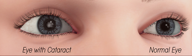
Cataracts
Cataracts

The human eye lens is composed of crystalline proteins. These proteins act to keep the lens clear. Shortly after you are born, all the fiber cells in the eye lose the ability to make new proteins, or to discard old proteins.
Cataracts can develop if the proteins become disrupted, by misfolding and then clump together, and break down, which is common with advancing age.
This clump is known as a cataract. Over time, the cataract gets worse and makes more of your lens cloudy.
Risk factors and causes include:
- Aging, stress, or hereditary effects.
- Eye surgery, such as retinal surgery.
- Vegetable oils, as they oxidize all the cells.
- Certain medications, such as prednisone steroids.
- Foods like sugar, grains, starchy fruits/vegetables.
- Health factors like diabetes, oxidative stress in cells.
- Smoking, alcohol (red wine OK), smoke> cell oxidation.
- Glycation, a natural process when excess sugar molecules in our skin fibers adhere to our skin’s collagen and elastin proteins.
Symptoms:
- cataracts cause images to appear blurry, like looking through a foggy window;
- difficulty seeing at night, less light in retina;
- changes in crystalline structure in lens, causing frequent changes in prescription glasses;
- changes in perception of colors;
- increased sensitivity to light;
- overlapping double/triple images;
- loss of contrast.
Unrelated symptoms:
- floaters in vision;
- pain or discomfort in the eye;
- film on surface of eye;
- fluctuation in vision (dry eye disease).

Eyedrops for Cataract Treatment
Crystallins are the major component of fiber cells, which form the eyes’ lenses, and the unique properties of these cells make them particularly susceptible to damage.
In order for our lenses to function well, this permanent, finite reservoir of crystallins must maintain both the transparency of fiber cells and their flexibility, as the eyes’ muscles constantly stretch and relax the lens to allow us to focus on objects at different distances.
The crystallins accomplish these duties with the help of aptly named proteins known as chaperones, which act “like antifreeze,” keeping crystallins soluble in a delicate equilibrium that’s in place for decades and decades.
They help prevent the clumping of proteins, or aggregation of insoluble amyloids (clumped-together proteins), that cause cataracts, but crystallins can become overwhelmed as we age.
Studies on human lens tissue showed that Lanosterol, which belongs to a group of chemical compounds called sterols, reverses protein aggregation in cataracts, and transparency increased. However, lanosterol was not water-soluble enough to be included in an eye drop solution and had to be injected into the eye.
In laboratory dish tests, the new team confirmed that sterol compound 29 significantly stabilized crystallins and prevented them from forming amyloids. They also found that compound 29 dissolved amyloids that had already formed.
Through these experiments, new researchers are starting to understand the mechanism in detail. They know where compound 29 binds, and are now beginning to know exactly what it is doing. A company called ViewPoint Therapeutics, is actively developing compound 29 for human use.
If you look at an electron micrograph of the protein aggregates that cause cataracts, you are hard-pressed to tell them apart from those that cause Alzheimer’s, Parkinson’s, or Huntington’s diseases.
Studying cataracts, enabled researchers to benchmark certain technologies and show by proof-of-concept that these technologies could also be used in nervous system diseases.
A cataract might appear cloudy to an optometrist, yet the patient says he can see well. On the other hand, another patient may have a clearer-looking cataract, yet complains about poor eyesight.

The patented product formula is guaranteed
by the Can-C hologram on all IVP eye drops boxes.
A product was developed in Russia, by using a compound called N-acetylcarnosine in eye drops to treat cataracts.
Topical application of carnosine onto the ocular surface does not result in penetration into the eye. Hence, a vehicle has been developed, called Nâ€acetylcarnosine (NAC). When NAC is instilled onto the eye it penetrates the anterior chamber of the eye through the cornea. Subsequent metabolism of NAC within the eye produces Lâ€carnosine, the active drug.
Lâ€carnosine has been shown to have an antioxidant effect on the cataractous lens. See Babizhayev 1989. Consequently, it is possible that topical administration of NAC could lead to a reduction in cataract by either slowing down the progression of the cataract or reversing the cataractous change.
Innovative Vision Products (IVP) is the patent holder and the only maker of the clinically proven N-Acetylcarnosine formula Can-C™, and is maintained as raw product for formula production control in a Japanese cGMP facility. To be sure you buy the patented product, the approved manufacturer Profound Products shows the Can-C hologram on the front of every box of their Can-C eye drops.
If the above is not the solution, lens replacement through surgery is right now the only other alternative.
Surgery
Cataract surgery is a refractive surgery, the surgeon can change your prescription after removal of the natural, fluid lens which had a certain power. The new acrylic lens has its own power and no auto focus anymore. After optimal vision, the secondary objective of the surgery is the type of prescription to determine what to see clear without glasses and when to use glasses.
Cataract surgery involves extracting the cataract and fluid lens material, and inserting a new, clear acrylic lens implant. The type of lens is based on clarity of focus at close-up, medium range, and distance. Crucial for treatment success is choosing the correct intraocular lens (IOL) power.
With glasses a minus sign before the number indicates that you are nearsighted, while a plus sign means that you’re farsighted. Lens power is measured in diopters. The higher the number, the stronger the prescription. For example, “-5.00” written under sphere means that you are very nearsighted and need a five diopter correction.

The refractive power of the human eye is dependent on three factors, the power of the cornea, the power of the lens (and where it will sit in the eye) and the axial length of the eye.
One of the most important parameters in IOL calculation, particularly in non-syndromic myopia, is axial length (AL). AL is a combination of anterior chamber depth (ACD), lens thickness and vitreous chamber depth and can change the IOL power by up to 2.5 to 3 times.

Optical methods use partial coherence laser tomography, and the interferometry principle to calculate distance from the cornea to the retina. Once measurements are made, the power of the replacement intraocular lens can be calculated.

The procedure is usually performed under local anesthesia on an outpatient basis, taking less than an hour in most cases.

During cataract surgery, an ophthalmologist breaks up the cataract by ultrasound or laser. Then removes the debris and inserts a mono-, or multi-focus lens (Ø 6mm) implant.

ZEISS AT LISA tri 839MP IOL multi-focal lens was found to provide a significantly better far distance retinal image quality than the PanOptix at both 3.0 and 4.0 mm pupil diameter.
It is the only IOL in the study providing a significantly higher mean postoperative value of far distance retinal image quality than the monofocal spherical control group.
The far distance retinal image quality is of great importance for neuroadaptation, as reduced image quality with blurred vision limits the neuroadaptation process.
The TECNIS® Eyhance Multifocal IOL can boost your vision in all light conditions, also while driving at night.

The PANOPTIX multi-focal causes glare/halo’s around night lights. Other brands are SYNERGY, VIVITY, SYMFONY.

Excellent equipment for intra-operative eye-measurements is the ORA Wavefront Aberrometer, a camera attached to the surgeon’s microscope allowing him/her to take pictures and do fine measurements of the eye after removing the cloudy lens, but before placing the new lens, to confirm or improve the choice of lens power.


PANOPTIX is the best lens for far-mid-near vision. Disadvantage is halo of lights at night. To resolve this the 2nd eye should have a monofocal lens that allows clear vision at far and mid, but requires glasses to see near, such as the Vivity lens. Thus, a combo of both gives the best results overall: Our brains can integrate the 2 different lens optics to see well at all distances day and night.

Multifocal intraocular lenses go further and allow you to see well at all distances. However, this type of intraocular lens requires a visual neuro-adaptation. In other words, the brain must adapt to the new focuses created by the lens.
At Vistaláser, they always check with contact lenses that the patient will adapt well before performing the implant. To obtain the best result it is important to carry out a good case study before the operation. In this way, they can be sure of choosing the most appropriate intraocular lenses for the patient, such as the Finevision lenses from Bausch & Lomb.
A Light Adjustable Lens (LAL), allows the surgeon to adjust the power of the lens by means of LIGHT, after the eye is healed from cataract surgery. However, initially the light of the sun can change the shape of the lens at random configuration. Thus, very special UV-protected sunglasses have to be worn at all times, until all the power lock-in treatments (5 additional visits) are done.

Recovery from cataract surgery normally takes around 8 weeks. During this period, a series of follow-up visits with the surgeon or your own Optometrist are required, to check the eyes to make sure they aren’t developing an infection and are healing properly.
From a few months to over a few years after surgery, about 50% will develop cloudiness in the capsule, which is called Posterior Capsule Opacification (PCO). A neodymium yag laser can remove the tiny epithelial cells, that created the original natural lens, which the surgeon could not remove, and they keep regenerating infinitely, and so proliferate after surgery, and cause hazyniss or glare.
Macular degeneration
This is a disorder that affects the retina, the light-sensitive lining at the back of the eye where images are focused. The macula -the area on the retina responsible for sharp central vision- deteriorates, causing blurred vision. This can cause difficulty reading and, for some, a blurry or blind spot in the central area of vision.
The most common form of age-related macular degeneration is known as non-exudative, or the "dry" form, in which vision loss usually progresses slowly. More rapid and severe vision loss comes from exudative, or the "wet" form, of macular degeneration. In the wet form, abnormal blood vessels develop under the macula and leak fluid and blood.
Glaucoma
This causes damage to the optic nerve, often due to increasing internal pressure in the eye because of problems with the flow or drainage of fluid within the eye. It can also occur when the internal pressure of the eye does not increase (normal-tension glaucoma), but there is not enough blood flow to the optic nerve. There are no early symptoms in the most common form of glaucoma, but the first signs of damage are defects in peripheral vision and difficulty with night vision. If diagnosed early, it can be treated with drugs, or sometimes surgery can minimize vision loss.

Examples of Refractive Error:
Nearsightedness: or myopia, is a condition in which nearby objects are seen clearly, but distant ones are blurred. Nearsightedness can be inherited and is often discovered during childhood. People that develop myopia in early adulthood usually do not develop high amounts of nearsightedness.
Farsightedness: or hyperopia (or hypermetropia), usually causes distant objects to be seen clearly, but close objects to appear blurred. Farsightedness often runs in families. When someone has higher levels of farsightedness, their distance vision may become blurry in addition to their near vision.
Astigmatism: occurs when the cornea is curved more in one direction, causing blurry vision at all distances, often occuring along with farsightedness or nearsightedness. Most people have small amounts of astigmatism. Larger amounts cause distortion in addition to blurry vision. Very high amounts of astigmatism causes difficulty with achieving 20/20 vision. Both a toric lens, LAL, and a multi-focal lens correct this plus a proper focus of far & near sight.

Presbyopia: this is the normal aging process of the lens of the eye, usually after age 40. It is the loss of elasticity of the lens that occurs with aging, causing difficulty focusing at close ranges. Scientists also believe that in addition to the loss of elasticity of the lens, the muscle that makes the lens change focus, called the ciliary body, also begins to not work as well. Ambyopia is when one eye is weaker than the other.
Dry eye disease: There are 3 ways that dry eyes can affect the cataract surgery:
- To best calculate what is the best power of the lens implant to be used, the surgeon will do measurements before surgery, such as the front curvature of the cornea. Dry eyes often damage the top epithelial cell layers, which will affect the accuracy, consistency of these measurements. A healthy cornea is key;
- The cornea will heal slower. The vision outcome could be worse than it was;
- Clean eyes: Blepharitis is a very mild but chronic bacterial lid infection caused by its waste products that then fall onto the surface of the eye and cause inflammation, called Endophthamitis, which could result in dry eyes and vision loss.
Others: lazy eye, head trauma, retinal problems, remnants of current/past life emotional trauma.

DIET
Adding grass-fed beef or egg-yolk, currants, selenium, or oculotrophin, powerful vitamins, antioxidants like anthocyanins that relax the ciliary muscle, e.g. in red cabbage, and in aronia and other berries, and minerals to your Keto-type diet will improve your vision and eye health:
Lutein & Zeaxanthin
Many studies show that lutein and zeaxanthin reduce the risk of chronic eye diseases. The more lutein and zeaxanthin the lower the risk for developing new cataracts. Dark green leafy vegetables are the primary source of lutein and zeaxanthin, as well as other colorful fruits and vegetables like broccoli, corn, peas, persimmons and tangerines.
Vitamin C
Scientific evidence suggests water-soluble vitamin B1, B2, & C lowers the risk of developing cataracts and when taken in combination with other essential nutrients, it can slow the progression of age-related macular degeneration and visual acuity loss. For your daily dose, try incorporating oranges, grapefruit, strawberries, papaya, green peppers and tomatoes into your diet.
Vitamin E
Vitamin E protects cells in the eyes from unstable molecules called free radicals, which break down healthy tissue. Good food sources of fat-soluble Vitamin E, A, D, K2, include vegetable oils (including safflower and corn oil), nuts, wheat germ and sweet potatoes.
Essential fatty acids
Omega-3 fatty acids are important for proper visual development and retinal function. Salmon, tuna and other cold-water fish are the best sources of omega-3 fatty acids and can help reduce inflammation, enhance tear production and support the eye’s oily outer layer.
Zinc
Zinc plays a vital role in bringing vitamin A from the liver to the retina in order to produce melanin, a protective pigment in the eyes. Impaired vision, such as poor night vision and cloudy cataracts, has been linked to zinc deficiency. For natural dietary sources of zinc, try red meat, oysters and other shellfish, and nuts and seeds.